Generated with Monsha
Mastering Mitosis: The Mechanics and Regulation of Cell Division
Anything · Grade 12 · English
Mastering Mitosis: The Mechanics and Regulation of Cell Division
Introduction
In the study of Grade 12 Biology, mitosis is recognized not just as "cell division," but as a highly regulated and intricate orchestration of molecular events. It is the process by which a eukaryotic cell divides its nucleus into two identical daughter nuclei, ensuring genetic continuity. This resource explores the phases of the cell cycle, the mechanics of mitosis, and the complex regulatory systems that govern these transitions.
1. The Cell Cycle: Preparation for Division
A cell spends the majority of its life in Interphase, a period of high metabolic activity where it prepares for the mitotic phase. Interphase is divided into three distinct stages:
- Phase (First Gap): The cell increases in size and synthesizes enzymes and proteins required for DNA replication.
- Phase (Synthesis): The cell’s DNA is replicated. Each chromosome now consists of two identical sister chromatids joined at a centromere.
- Phase (Second Gap): The cell continues to grow and produces the proteins necessary for the mitotic spindle. This is the final preparation stage before the M phase begins.

2. The Phases of Mitosis
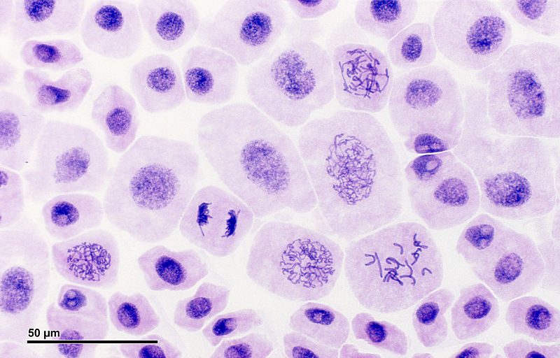
Mitosis is a continuous process traditionally divided into five stages for ease of study:
Prophase
Chromatin fibers condense into discrete chromosomes visible under a light microscope. The nucleolus disappears, and the centrosomes begin to move to opposite poles, initiating the formation of the mitotic spindle.
Prometaphase
The nuclear envelope fragments, allowing spindle microtubules to enter the nuclear area. These microtubules attach to the kinetochores—specialized protein structures located at the centromeres of each sister chromatid.
Metaphase
The centrosomes are now at opposite poles of the cell. The chromosomes convene at the metaphase plate, an imaginary plane equidistant between the spindle's two poles. For every chromosome, the kinetochores of the sister chromatids are attached to microtubules coming from opposite poles.
Anaphase
The shortest stage of mitosis. The cohesin proteins holding sister chromatids together are cleaved, allowing them to separate. The daughter chromosomes move toward opposite ends of the cell as their kinetochore microtubules shorten.
Telophase
Two daughter nuclei form in the cell. Nuclear envelopes arise from the fragments of the parent cell's nuclear envelope and other portions of the endomembrane system. The chromosomes become less condensed, and the spindle is depolymerized.
3. Cytokinesis: The Final Split
While mitosis is the division of the nucleus, cytokinesis is the division of the cytoplasm. This process differs between animal and plant cells:
- Animal Cells: A contractile ring of actin and myosin filaments forms a cleavage furrow, pinching the cell in two like a drawstring.
- Plant Cells: Due to the rigid cell wall, a cleavage furrow is impossible. Instead, vesicles from the Golgi apparatus move to the middle of the cell and fuse to form a cell plate, which eventually becomes a new cell wall.
4. Regulation and Quality Control
At the Grade 12 level, understanding the control of the cell cycle is paramount. The cell uses a molecular control system to ensure division only occurs under favorable conditions.
Checkpoints
- Checkpoint: The "restriction point." If the cell receives a go-ahead signal, it usually completes the cycle. If not, it may exit the cycle and enter, a non-dividing state.
- Checkpoint: Ensures that DNA replication in thephase was successful and that the DNA is undamaged.
- M Checkpoint (Spindle Checkpoint): Occurs during metaphase. It ensures that all chromosomes are properly attached to spindle microtubules before anaphase begins.
Cyclins and CDKs
The cell cycle is regulated by two types of proteins:
- Cyclins: Proteins whose concentration fluctuates rhythmically during the cycle.
- Cyclin-Dependent Kinases (CDKs): Enzymes that are always present but are only active when attached to a specific cyclin.
For example, the MPF (Maturation-Promoting Factor) is a cyclin-CDK complex that triggers the cell's passage past the
checkpoint into the M phase. When the concentration of cyclin rises in
, it binds with CDK to form active MPF, which phosphorylates proteins to initiate mitosis.5. Biological Significance
- Growth and Development: Allows a single-celled zygote to grow into a multicellular organism.
- Tissue Repair: Replaces dead or damaged cells (e.g., skin regeneration).
- Asexual Reproduction: For many single-celled eukaryotes and some multicellular organisms, mitosis is the mechanism of reproduction, producing clones of the parent.