Generated with Monsha
Mitosis and the Eukaryotic Cell Cycle
Anything · Grade 12 · English
Mitosis and the Eukaryotic Cell Cycle
Introduction to the Cell Cycle
In eukaryotic organisms, the cell cycle is an ordered series of events involving cell growth and division that produces two new daughter cells. For somatic (body) cells, this process ensures that each new cell receives an identical set of genetic information. The cell cycle is broadly divided into two main phases: Interphase and the Mitotic (M) Phase.
1. Interphase: Preparation for Division
Interphase is the longest part of the cell cycle, during which the cell grows and replicates its DNA. It is divided into three distinct sub-phases:
- G1 Phase (First Gap): The cell undergoes rapid growth and carries out normal metabolic functions. It accumulates the building blocks of chromosomal DNA and the associated proteins, as well as enough energy reserves to complete the task of replicating each chromosome in the nucleus.
- S Phase (Synthesis of DNA): This is the most critical part of interphase. The cell replicates its entire genome. Each chromosome is duplicated to form two identical sister chromatids joined at a central region called the centromere. The centrosome is also duplicated during this phase.
- G2 Phase (Second Gap): The cell replenishes its energy stores and synthesizes the proteins necessary for chromosome manipulation. Some cell organelles are duplicated, and the cytoskeleton is dismantled to provide resources for the mitotic spindle.

2. The Mitotic (M) Phase
The M phase involves two major processes: Karyokinesis (nuclear division) and Cytokinesis (cytoplasmic division). Karyokinesis is divided into five stages for detailed study at the Grade 12 level.
Prophase
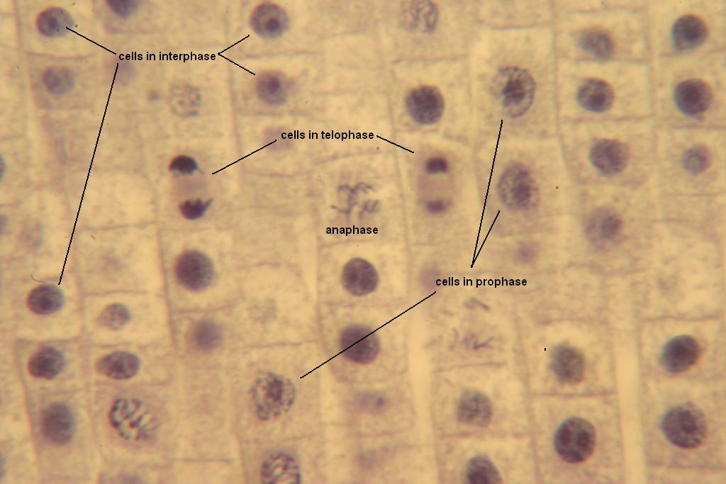
Chromatin fibers condense into tightly coiled chromosomes, visible under a light microscope. Each duplicated chromosome appears as two identical sister chromatids joined at their centromeres. The nucleolus disappears, and the mitotic spindle—composed of microtubules—begins to form between the centrosomes as they move to opposite poles of the cell.
Prometaphase
The nuclear envelope breaks down into small vesicles. Microtubules from the spindle reach the chromosomes and attach to kinetochores, specialized protein structures on the centromeres. These "kinetochore microtubules" begin to jerk the chromosomes back and forth.
Metaphase
The centrosomes are now at opposite poles of the cell. The chromosomes convene on the metaphase plate, an imaginary plane that is equidistant between the spindle’s two poles. The centromeres of all the chromosomes are aligned on this plate. For every chromosome, the kinetochores of the sister chromatids are attached to microtubules coming from opposite poles.
Anaphase
This is the shortest stage of mitosis. The cohesin proteins holding the sister chromatids together are cleaved, allowing the chromatids to separate. Each chromatid becomes a full-fledged chromosome. These daughter chromosomes move toward opposite ends of the cell as their kinetochore microtubules shorten. By the end of anaphase, the two ends of the cell have equivalent—and complete—collections of chromosomes.
Telophase
Two daughter nuclei form in the cell. Nuclear envelopes arise from the fragments of the parent cell's nuclear envelope and other portions of the endomembrane system. Nucleoli reappear. The chromosomes become less condensed, and the remaining spindle microtubules are depolymerized. Mitosis, the division of one nucleus into two genetically identical nuclei, is now complete.
3. Cytokinesis: The Final Split
While karyokinesis divides the genetic material, cytokinesis divides the cytoplasm and organelles to create two distinct cells. This process differs between animal and plant cells due to structural differences.
- In Animal Cells: A contractile ring of actin microfilaments and myosin proteins forms on the cytoplasmic side of the plasma membrane. This ring contracts, pulling the membrane inward and forming a cleavage furrow. The furrow deepens until the parent cell is pinched in two.
- In Plant Cells: Because of the rigid cell wall, a cleavage furrow cannot form. Instead, vesicles derived from the Golgi apparatus move along microtubules to the middle of the cell, where they fuse to produce a cell plate. The cell plate grows outward until its surrounding membrane fuses with the plasma membrane along the perimeter of the cell. A new cell wall then forms from the contents of the cell plate.
Significance of Mitosis
Mitosis is vital for three primary reasons:
- Growth: It allows a single-celled zygote to grow into a multicellular organism.
- Tissue Repair: It replaces damaged or aged cells in tissues.
- Asexual Reproduction: In many unicellular and some multicellular eukaryotes, mitosis is the primary means of reproduction, creating genetically identical offspring.